New BrIMA study confirms increased intraoperative success rates in breast surgery using the XEOS AURA 10
25th June 2026
JAMA Surgery has published the findings which features the mobile PET-CT specimen imager
A new BrIMA (Breast cancer Intraoperative Margin Assessment) study has been published in the world’s leading peer-reviewed surgical journal, JAMA Surgery. The prospective, multicenter clinical trial demonstrated that the specimen images obtained by the XEOS AURA 10 PET-CT system significantly improved surgeons’ ability to identify and address positive margins during breast-conserving surgery, providing actionable information at the point where surgical decisions are made.
As the largest evaluation of its type conducted to date, BrIMA provides important new evidence supporting the role of intraoperative molecular imaging in surgical decision-making.
Positive margins remain one of the most persistent challenges in breast-conserving surgery, occurring in approximately 12–30% of procedures and representing the leading cause of reoperation. When residual cancer is discovered only after postoperative pathology review, patients may require a second surgery, resulting in additional physical and emotional distress, delayed treatment, increased healthcare costs, and compromised cosmetic outcomes.
Key findings
- In patients with invasive ductal carcinoma (IDC), the most common form of breast cancer, intraoperative success rates increased from 83.3% without intraoperative margin assessment to 95.2% with specimen PET-CT imaging.
- Across all breast cancer subtypes included in the study, success rates improved from 76.4% without intraoperative margin assessment to 91.9% with specimen PET-CT.
- No device-related adverse events were reported, and strong agreement was observed among surgeons interpreting specimen PET-CT images.
"The most important finding from BrIMA is that surgeons gained additional insight at the exact moment they needed to make a decision," said Vincent Keereman, Founder and CEO of XEOS. "Too often, positive margins are discovered only after surgery is complete and the opportunity to act has passed. BrIMA demonstrates that specimen PET-CT can provide actionable information while the patient is still in the operating room, enabling surgeons to make more informed decisions when corrective action is still possible."
Actionable results in 10 minutes
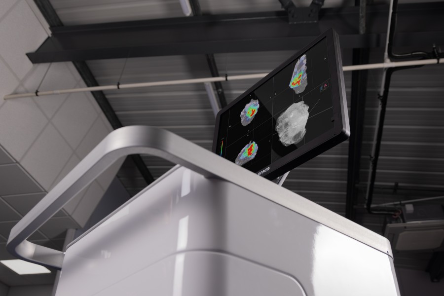
The AURA 10 specimen PET-CT system differs from conventional approaches that evaluate only portions of a specimen or provide limited two-dimensional information. By combining molecular and three-dimensional imaging in a single scan, AURA 10 provides high-resolution visualisation of the entire excised specimen. The system is designed for use directly within the surgical workflow, delivers results in approximately 10 minutes, and does not require specimen transport outside the operating room.
“AURA 10 has the potential to help surgeons identify positive margins …”
“The BrIMA results demonstrate that specimen PET-CT can provide reliable intraoperative visualisation across multiple breast cancer subtypes while integrating effectively into routine surgical practice,” said Menekse Göker, MD, primary author of the publication and breast surgeon at Ghent University Hospital. “By providing additional information during surgery, AURA 10 has the potential to help surgeons identify positive margins that might otherwise only be discovered after postoperative pathology review, allowing corrective action while the patient remains in the operating room.”
Breast-conserving surgery remains the standard treatment for many women diagnosed with early-stage breast cancer. As healthcare systems increasingly focus on reducing reoperations, improving patient outcomes, and optimizing surgical efficiency, technologies that provide more complete intraoperative information may play an increasingly important role in surgical oncology. The BrIMA findings further support the potential role of specimen PET-CT in helping surgeons make more informed intraoperative decisions and may provide a foundation for future applications across additional oncologic procedures.
About the BrIMA study
BrIMA (ClinicalTrials.gov Identifier: NCT04999917) was a prospective, multicenter, interventional clinical study conducted between June 2022 and March 2025. The study analyzed 148 patients undergoing breast-conserving surgery at six breast cancer centers in Belgium, Germany, and Italy. The primary endpoint evaluated the success rate of specimen PET-CT imaging in addressing positive margins in patients with invasive ductal carcinoma. The study demonstrated statistically significant improvements in intraoperative margin assessment and surgical success rates when specimen PET-CT was incorporated into routine surgical workflow.
Find out more
You can learn more about the XEOS AURA 10 and access the full publication by clicking the button below.