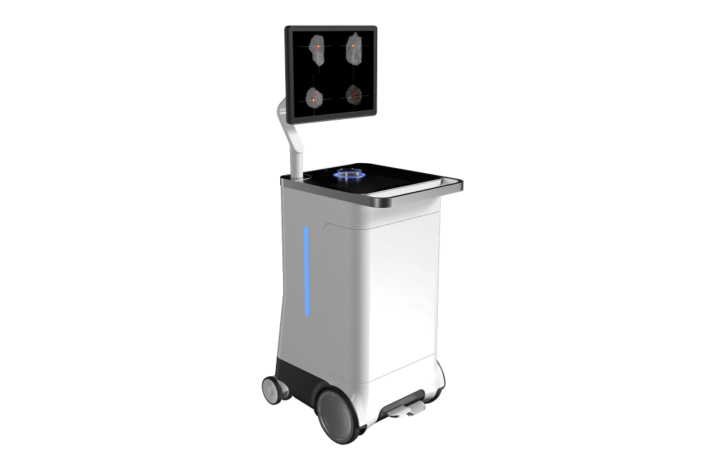
The first PET-CT specimen imager for the OR

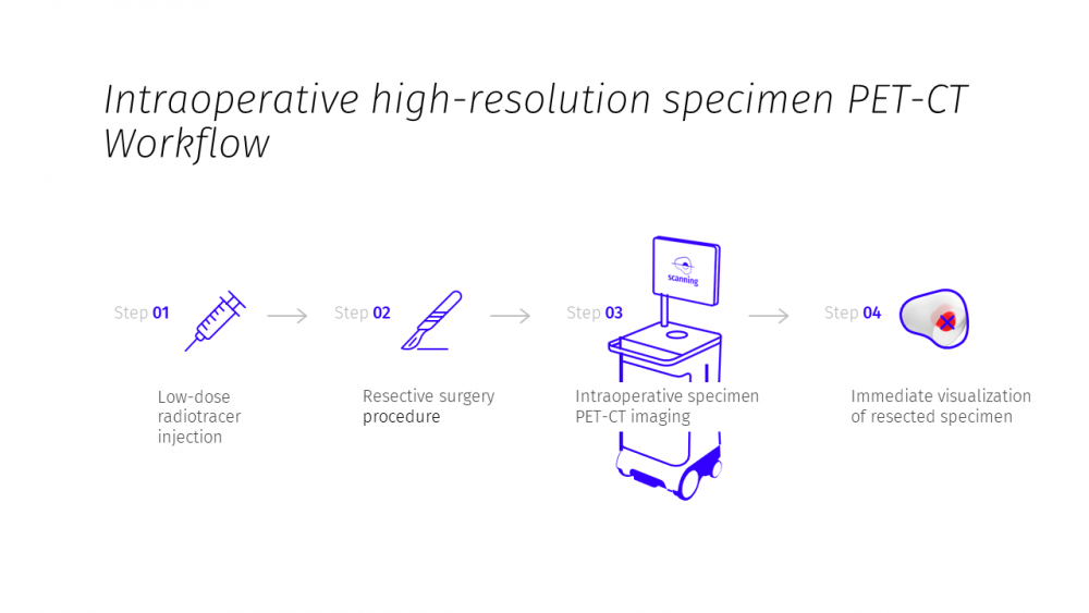
In surgical oncology, such as breast conserving surgery, rapid assessment of the excised specimen is of utmost importance. You want to be confident that your surgery reached its goal of resecting the target lesion, so that your patients recovery can be promoted.

AURA 10 has made molecular imaging available in the OR for the first time. As a result, surgeons and imaging specialists have never been able to perform specimen imaging with so much confidence. Ultimately, your patients will be the ones who benefit.

Benefits of intra-operative specimen imaging

Less chance of resurgery

The AURA 10 combines the picomolar sensitivity of PET with high-precision 3D image reconstruction. This gives you all the imaging capabilities you need to image the targeted tissue in a single procedure, to close your patient with more confidence, and to reduce the probability of resurgery.

More peace of mind for your patients

Using the best available intraoperative imaging technology will significantly decrease the anxiety level of the patient. This stress reduction will contribute to your patient’s recovery.

Reduced anesthesia time

A more streamlined workflow and real-time collaboration with your radiology department can shorten the procedure. This may also reduce anesthesia time for your patient and decrease the risk of postoperative complications.

Better cosmetic outcome

Intraoperative imaging can help you to remove less tissue in the initial surgery and to obtain a better cosmetic result.

Transforming surgical oncology

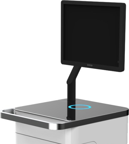
With the AURA 10 PET-CT Imager, XEOS is transforming mobile specimen radiography, allowing you to operate with more clarity. AURA 10 brings increased precision to the OR by integrating PET molecular imaging and 3D tomography acquisition capabilities into a compact and ergonomic cart-based system. This makes it possible for the first time to obtain reference image quality in your procedure room within minutes after excision.

Key features of AURA 10

Powerful medical imaging processing

Once acquired, you want your PET-CT images to be reconstructed and visualized fast and accurately. With the AURAFLOW visualisation and reconstruction engine, your clip verification images are available within 30 seconds, and the reconstructed high-resolution CT after only 3 minutes. Metal artifact correction avoids any image disturbance due to clips in the CT image. Your PET images are available within 10 minutes after data acquisition.

AURAFLOW allows you to zoom in on your co-registered PET and CT images and to make annotations. You can also measure, scroll through slices, rotate the image, adapt the window level, and much more. The DICOM interface assures data compatibility.

Save procedure time

Transporting your specimen to the radiology department and receiving diagnostic results takes time. Meanwhile, you need to wait and lose valuable minutes before closing your patient with more confidence. With the AURA 10 specimen imager, there is no transport required. Just obtain reference image quality at the point of surgery and upload the images to your PACS, so your imaging specialist can view them right away. This saves valuable procedure time.

High-precision 3D data

2D images obtained with conventional specimen radiography are valuable, but the limited sensitivity of this technology still makes it hard to accurately localize tumor cells in the specimen. The AURA 10 offers more clarity and confidence, because it provides high-precision 3D data that allows you and your imaging experts to verify samples inside the specimen with more certainty. The AURA 10 clearly highlights the radiotracer uptake of the tissue and indicates its location, shape and size inside the harvested specimen.

Integrate seamlessly into your OR

The compact AURA 10 imager has a high-quality finish and will seamlessly integrate into your OR environment. It’s very mobile, so you are free to give it the most suited place in your procedure room or to share it with other surgical oncologists. The AURA 10 starts up in less than 10 minutes and the solid shielding of the x-ray source will offer your surgical staff the highest protection during use.

Compact unit

The AURA 10 offers high-performance PET-CT molecular imaging capabilities, all engineered into the smallest, most practical form factor. It’s highly mobile for use in and out of your operating room, and while offering a similar level of precision, it only comes at the fraction of the cost of a conventional, large-scale PET-CT scanner.

Ergonomic design

The AURA 10 is easy and intuitive and allows you to fully focus on your patient. Your surgical staff will appreciate the unit’s compact design, curved transportation handle, easy brake system, and motorized top-load specimen receptacle. The medical-grade display monitor can be tilted and swiveled to give you more viewing comfort.

Safe for your staff

An LED ring around the top-load specimen receptacle will indicate proper tray insertion, specimen position and closure. A vertical LED bar on both sides of the unit shows when the x-ray acquisition is taking place and informs you about the progress of the image acquisition. The x-ray source is appropriately shielded, which avoids exposure to the surgical staff. The PET scanner only requires a minimal radiotracer marker dose, and fully complies to Radiation Protection & Safety of Radiation Sources standards.

Practical interface

The integrated touch-control display and trackpad enable you to instantly set up the image acquisition and obtain the required views with little image manipulation. Uploading the images to your PACS is easy and fast. The intuitive user interface enables you to simply link the acquired images to the patient and procedure data within the PACS system.

More clarity for more patients

The AURA 10 PET-CT Imager offers surgical oncologists the opportunity to assess resected specimens intraoperatively with more clarity and for an increasing number of applications and patients. Surgeons can now operate with more confidence, but also speed up procedure time, reduce anesthesia time, and improve the chances of recovery.

The AURA 10 can be used for a diverse range of applications, including:

Breast conserving surgery

Surgical oncologists rely on the AURA 10 PET CT imager to obtain high-precision images of the breast specimen while minimizing the disruption of the surgical workflow.

Breast surgeons need high-precision images of resected specimens to reduce the risk of resurgery and improve the cosmetic outcome as much as possible. Unfortunately, today’s conventional intraoperative techniques for margin assessment are either labor-intensive or only offer limited functionality.

The new AURA PET-CT imager however makes use of 18F-FDG radiotracer to provide surgeons with more clarity on breast specimens in a matter of minutes. This reduces the operating time, increases chances of recovery, and improves cosmetic results.

Prostate cancer surgery

Urology surgeons can include the AURA 10 PET-CT imager in their procedures to assess negative margins intraoperatively with more certainty. This way, they can conduct nerve-sparing prostatectomies with a higher chance of success.

A clear picture of the resection margins is crucial if you want to reduce the chances of reoperation or adjuvant therapies like radiotherapy or chemotherapy. To date, frozen sections are still most common in prostate cancer surgery. However, this technique requires a long intraoperative evaluation and anesthesia period (up to 30 minutes), which is not beneficial for the patient.

The AURA PET-CT imager on the other hand leverages the power of PSMA PET and delivers a high-precision image of the specimen in a matter of minutes. This speeds up procedure time, reduces anesthesia time, and improves patient outcomes.

Head & neck cancer surgery

During head and neck cancer surgery, the AURA 10 PET-CT imager can give a clear, accurate view of the resected specimen. This enables surgeons to assess deep margins, even in the smallest oral cancers.

For head and neck cancer surgery, assessing surgical margins and making the right decisions is a complex process. The number of positive margins is typically high, compared to other types of cancer surgery, and until now, the possibilities for intraoperative assessments have been limited. Frozen sections are widely used for intraoperative assessment, but unfortunately, they prolong the operation time significantly.

The AURA PET-CT imager however makes use of 18F-FDG radiotracer to provide surgeons with more clarity on neck or head specimen in a matter of minutes. The result is a shorter operating time and less risk of resurgery or adjuvant therapies for the patient.

XEOS AURA 10 existing users

Click on the tabs below to find out more about current customers and how they are using the AURA 10 in their hospitals.

Ghent University Hospital, Belgium

Reducing resurgeries and salvage radiotherapies in breast conserving surgery

Based in Ghent, Belgium, XEOS have developed excellent collaborations with local hospitals. In a new video, Dr. Menekse Göker of Ghent University Hospital demonstrates the AURA 10’s clinical workflow in a patient diagnosed with invasive ductal carcinoma who underwent surgery with a sentinel lymph node resection.

Find out how the AURA 10 allowed Dr. Göker to visualise the tumour in real time in the operating room, which lead to the identification of a positive resection margin after the initial sentinel lymph node resection.

READ MORE

Documents

Sorry, there are no documents available for this product.

The LabLogic Group will always treat your details with the utmost care and will never sell them to third party companies.

Please choose one of the following:


Previous
Next